
How Thick Should the Lining Be for Embryo Transfer?

Key Takeaways
Many clinics aim for a lining of about 7 mm before embryo transfer. But the number is not read on its own. I also check for fluid in the uterus and the timing of progesterone. I consider how the lining looks, the embryo, the type of transfer and any past problems with the uterus. Together, these help me decide whether to go ahead, change the plan or look further.
Key evidence: Liu et al. — Thin lining and IVF outcomes (Human Reproduction, 2018) Genovese et al. — Endometrial thickness in euploid FET (Human Reproduction, 2025) ESHRE — Recurrent implantation failure good practice (2023)
On this page
- What we measure before transfer
- Is 7 mm the magic number?
- What if the lining is 6–7 mm?
- Can the endometrium be too thick?
- Pattern, fluid, and timing
- Why some linings stay thin
- What we can do when the lining is thin
- When postponing transfer may be considered
- Frequently asked questions for Dr. Aksoy
- Takeaway
- Sources
Endometrial thickness is one of the numbers patients remember most clearly before an embryo transfer. I understand why. After weeks, sometimes months, of treatment, a single ultrasound measurement can feel as if it decides the whole cycle.
In my practice, I do not look at that number in isolation. A lining of 6.8 mm is not the same story in every patient. A lining of 9 mm is not automatically reassuring if there is fluid in the cavity or if progesterone timing is wrong. The question is not only “How thick is the endometrium?” It is “Is this endometrium ready for this embryo, in this cycle?”
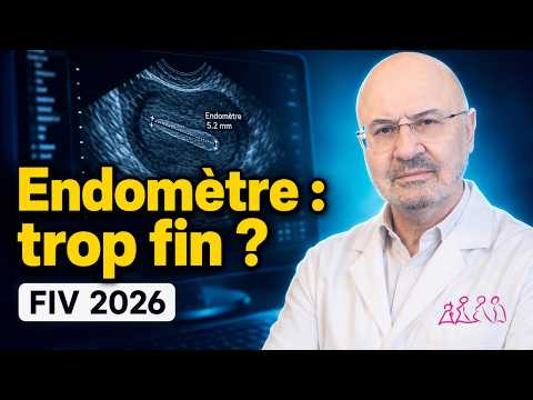
(Note: this video was recorded in French. English audio dubbing and subtitles are available — select your preferred audio track and subtitles in the YouTube player settings.)
What We Measure Before Transfer
Endometrial thickness is measured on transvaginal ultrasound in the midsagittal uterine plane, at the thickest part of the endometrium. The maximal double-layer thickness is measured perpendicular to the endometrial midline, excluding any fluid within the cavity (ISUOG consensus, 2026). The timing of the measurement relative to ovulation, trigger, or the start of progesterone is also recorded.
Very small decimal differences can sometimes reflect measurement technique, image quality, or observer variation rather than a clinically meaningful biological change. A reading of 6.8 mm is therefore not categorically different from 7.0 mm once the full scan and clinical context are considered.
Before transfer, I also pay attention to:
- endometrial appearance,
- whether there is fluid inside the uterine cavity,
- the timing of progesterone exposure,
- the type of cycle: fresh, natural frozen, modified natural, or programmed,
- and the patient’s uterine history.
Is 7 mm the Magic Number?
Many clinics use about 7 mm as a practical threshold before embryo transfer, especially in frozen embryo transfer cycles. Some use 7.5 or 8 mm. This is not because a pregnancy cannot happen below that number. It is because several studies show lower pregnancy or live birth rates when the lining is very thin.
A large cohort study by Liu and colleagues, covering more than 40,000 transfers, suggests that outcomes tend to decline below about 8 mm in fresh transfers and below about 7 mm in frozen transfers. An older meta-analysis by Kasius and colleagues also found that clinical pregnancy rates were lower when the endometrium was below 7 mm. The CFAS thin-endometrium guideline likewise treats thickness under about 7–8 mm as clinically relevant for counselling, while noting that cut-offs vary across studies.
But there is an important nuance. Endometrial thickness is a marker, not a verdict. In a large 2025 study of 30,676 single euploid frozen embryo transfers, thickness alone was a relatively weak overall predictor of live birth once embryo genetics were controlled. Even so, linings under 7 mm were still associated with lower live-birth odds in programmed and modified-natural cycles; the association was not significant in true natural cycles. These were retrospective associations, not proof that cancelling or delaying a transfer below a particular measurement improves the cumulative chance of live birth. Thresholds also varied between centres and cycle types, and the study included only cycles that reached transfer.
A 2025 SART CORS analysis by Schmiech and colleagues, covering more than 244,000 cycles, found an association between thickness and live birth up to about 9 mm. The authors cautioned that the data should not be used to withhold transfer below a particular thickness. A second 2025 analysis of the same registry by Gingold and colleagues found that live birth rates kept rising slightly up to about 12 mm. The two analyses reach somewhat different cut-offs, but both point to a gradual change rather than a sharp line.
What If the Lining Is 6-7 mm?
This is the gray zone where judgment matters.
If the lining is 6.5 or 6.8 mm, I do not automatically cancel the transfer. I ask a more practical set of questions:
- Is the lining still growing?
- Does it look regular?
- Is there any fluid in the cavity?
- Is this the only available embryo—particularly a euploid embryo—or are several embryos available?
- Has this patient had repeated thin lining despite different protocols?
- Are we in a natural, modified natural, or programmed cycle?
- Have there been previous uterine surgeries, curettage, a documented or clinically suspected intrauterine infection, or suspicion of adhesions?
Sometimes the best decision is to continue. Sometimes it is wiser to extend estrogen, change the route of medication, repeat the scan, or prepare a different cycle. When this is the only available embryo, I may take a more conservative approach. I also explain that thinner linings are linked with lower pregnancy and live-birth rates on average, but one decimal is not a rule.
How long I extend estrogen, and when I change the cycle type
In a programmed frozen embryo transfer, if the lining measures 6–7 mm on the planned day, I do not cancel straight away. If progesterone has not yet started, I usually extend estrogen by another 4–7 days, changing the dose or route if needed, and then measure again. In practice, for most patients I do not take total estrogen use much beyond 21–28 days, because there is no strong evidence that prolonging it past a certain point thickens the lining further. One cohort study of about 1,400 frozen blastocyst transfers also linked more than 28 days of estrogen before transfer with lower live birth rates (Bourdon et al., 2018).
I consider switching to a natural or modified natural cycle particularly for women who ovulate regularly and whose lining stays thin again and again in programmed cycles despite adequate estrogen. If I know the lining grew better in earlier natural or stimulated cycles, that decision becomes stronger. If, on the other hand, the lining stays stuck at around 5–6 mm, I first want to be sure there are no intrauterine adhesions or other problems in the uterine cavity. For me, the deciding factor is not a single millimetre but how the patient’s own endometrium behaves from one cycle to the next.
Can the Endometrium Be Too Thick?
Patients often worry about a lining that is “too thick.” In routine embryo transfer practice, the bigger concern is usually a persistently thin lining, not a moderately thick one.
A thick lining may still be acceptable if it looks regular and the cavity is clean. If it is unusually thick, irregular, or associated with abnormal bleeding, polyps, hyperplasia, retained tissue, or an unsynchronized hormonal response may need to be ruled out before transfer. A smooth 12 mm lining is not the same as an irregular 12 mm lining with suspicious findings.
Pattern, Fluid, and Timing
Before progesterone, a trilaminar pattern is commonly documented and may be clinically reassuring, but on its own it does not guarantee receptivity or live birth. After progesterone begins, the lining normally becomes more homogeneous, so the timing of the scan matters.
Fluid in the uterine cavity needs the same care. A small amount of fluid seen earlier in the cycle may resolve before transfer. In one cohort, fluid that had cleared by the day of transfer did not lower live birth rates (Nguyen et al., 2023). Fluid still present at or close to transfer is more concerning: in a meta-analysis of nine studies, fluid at the time of transfer was linked with lower odds of pregnancy (Kathuria et al., 2023). In a 2026 study of euploid transfers, persistent fluid was linked with a live birth rate of about 38%, compared with 59% without fluid; even so, cancelling for it did not clearly improve the chance at the next attempt (Patel et al., 2026). So I first look for the cause, including possible hydrosalpinx or uterine/cervical factors.
Progesterone timing remains a key decision point. In a frozen embryo transfer, the embryo stage and the number of progesterone days must match.
You can read more about timing and protocol choice in our guide to endometrial preparation for frozen embryo transfer.
Why Some Linings Stay Thin
A thin endometrium can happen for several reasons. Sometimes it is simply how that cycle behaves. Sometimes there is a history behind it.
Factors that may contribute include:
- previous curettage, uterine surgery, or intrauterine adhesions,
- inadequate hormone exposure or absorption in a programmed cycle,
- cycle-to-cycle variation and a repeatedly limited endometrial response,
- and, less commonly, other uterine or systemic factors.
Chronic endometritis or tubal disease such as hydrosalpinx may be investigated when the history, ultrasound findings, cavity fluid, or repeated treatment outcomes create a specific clinical suspicion. A thin measurement alone does not point to them.
A thin lining is a clinical finding that deserves a careful investigation. It is not the patient’s fault.
What We Can Do When the Lining Is Thin
The first step is usually simple: review the preparation protocol.
In programmed cycles, I review estrogen dose, duration, route, adherence, and absorption. Extending exposure or using another route is sometimes considered, but no single estrogen route or combination has been shown to be universally superior for refractory thin endometrium or to reliably improve live birth. A natural or modified-natural protocol may be considered when ovulation is reliable, although it is not a guaranteed treatment for thin lining.
If the lining remains thin across repeated attempts, I consider whether we are missing a uterine factor, especially after curettage, uterine surgery, a documented or clinically suspected intrauterine infection, or a repeatedly unexplained thin response. For women with recurrent implantation failure whose lining stays thin despite adjusting the preparation, ESHRE guidance recommends reviewing the estradiol treatment and says hysteroscopy can be considered to rule out adhesions.
I do not offer PRP or G-CSF routinely. No solid evidence shows that they increase live births. ESHRE does not recommend their routine use in recurrent implantation failure, and CFAS found insufficient evidence that adjuvants reliably improve pregnancy or live-birth rates in thin endometrium. The ASRM 2026 committee opinion on recurrent implantation failure likewise finds insufficient evidence to support PRP or G-CSF. When a patient asks about them, I explain what they are: experimental, paid treatments with uncertain benefit and possible risks. Before that point, I check four things: the diagnosis, estrogen exposure, timing and the uterine cavity.
The same caution applies to procedures such as endometrial scratching.
When Postponing Transfer May Be Considered
In my practice, I postpone the transfer when the lining stays below 6 mm. Pregnancy is still possible below that level, but in a large US registry analysis live birth rates were clearly lower: about 31% below 6 mm, compared with 45% at 8–11.9 mm, across fresh and frozen transfers (Gingold et al., 2025). I may also consider postponing when:
- there is persistent fluid in the uterine cavity,
- the lining looks irregular or suspicious,
- progesterone timing has become uncertain,
- there is a strong suspicion of intrauterine adhesions, a polyp, or another clinically relevant uterine-cavity abnormality,
- or the patient has only one or very few available embryos—particularly a euploid embryo—and the cycle feels suboptimal.
Postponing is not failure. For a borderline thickness alone, however, evidence that postponement improves cumulative live birth is limited (Kasius et al., 2014; Schmiech et al., 2025); I also weigh the delay, cost and burden for the patient, and try to separate a meaningful finding from a small measurement difference that mainly creates anxiety.
Frequently Asked Questions for Dr. Aksoy
How thick should the lining be for embryo transfer?
There is no single “ideal” or mandatory thickness. In practice, 7 mm or more is considered reassuring, and 7.5 mm is a perfectly reasonable thickness for transfer. Recent data, however, show that the chance of success changes gradually with thickness rather than at a sharp threshold (Schmiech et al., SART CORS 2025; Pérez-Milán et al., meta-analysis 2025). That is why I do not decide on a transfer from the millimetre reading alone.
Can pregnancy happen with a 6 mm lining?
Yes. Pregnancy and live birth are possible with a 6 mm lining, and even with a thinner one. Average success may be lower than with a thicker lining (Liu et al., 2018), but that does not mean pregnancy cannot happen below 7 mm.
Can the lining be too thick for embryo transfer?
For IVF, there is no proven upper limit above which transfer should not be done. Large studies have not shown a fall in live birth rates even at 15 mm or more (Gingold et al., SART CORS 2025). If the lining is unexpectedly thick, irregular or uneven in texture, though, the real question is less the thickness itself than whether there is a polyp, hyperplasia or another problem in the uterine cavity.
Do ASRM or ESHRE guidelines set 7 mm as a minimum?
No. 7 mm is a practical threshold that clinics often use, but neither ASRM nor ESHRE has a mandatory rule that transfer should not take place below 7 mm. ESHRE’s ovarian stimulation guideline, updated in 2025, does not recommend routine monitoring of endometrial thickness, citing evidence that thickness on its own predicts pregnancy poorly (ESHRE, 2025 update; Kasius et al., 2014). A recent review also points out that the commonly used 7 mm threshold does not rest on strong evidence (Pirtea and Ata, 2026).
Is a thick lining on ultrasound a sign of pregnancy?
Not on its own. The lining can thicken after ovulation under the effect of progesterone, or because of the hormones used in treatment. After a transfer, too, pregnancy cannot be confirmed or ruled out from endometrial thickness; it is assessed with a β-hCG blood test and later by seeing the gestational sac on ultrasound.
Takeaway
Endometrial thickness matters. I measure it carefully, and I take a persistently thin lining seriously.
But I also tell patients not to let one number take over the whole story. The endometrium must be synchronized with the embryo, and a good transfer decision weighs the scan together with everything else we know about the cycle.
— Dr. Senai Aksoy
Sources
- Liu KE, Hartman M, Hartman A, et al. “The impact of a thin endometrial lining on fresh and frozen-thaw IVF outcomes: an analysis of over 40,000 embryo transfers.” Human Reproduction. 2018. PubMed
- Kasius A, et al. “Endometrial thickness and pregnancy rates after IVF: a systematic review and meta-analysis.” Human Reproduction Update. 2014. DOI
- Genovese H, et al. “Does endometrial thickness impact live birth rate following a frozen embryo transfer: outcomes of 30,676 euploid single embryo transfers.” Human Reproduction. 2025. Human Reproduction
- Schmiech K, Li M, Chen LX, et al. “Association of endometrial thickness with live birth rate: a study using the Society for Assisted Reproductive Technology Clinic Outcome Reporting System.” Fertility and Sterility. 2025. DOI
- Canadian Fertility and Andrology Society. “Management of thin endometrium in assisted reproduction: a clinical practice guideline.” Reproductive BioMedicine Online. 2019. DOI
- ESHRE Working Group on Recurrent Implantation Failure. “ESHRE good practice recommendations on recurrent implantation failure.” Human Reproduction Open. 2023. HROpen
- Bourdon M, et al. “Prolonged estrogen (E2) treatment prior to frozen-blastocyst transfer decreases the live birth rate.” Human Reproduction. 2018. DOI
- Gingold JA, et al. “Increasing endometrial thickness up to 12 mm is associated with increased odds of live birth among fresh and frozen-thawed autologous transfers with or without preimplantation genetic testing.” Fertility and Sterility. 2025. DOI
- Pérez-Milán F, et al. “Impact of endometrial thickness on reproductive outcome in fresh and frozen-thawed embryo transfer: systematic review and meta-analysis.” Ultrasound in Obstetrics & Gynecology. 2025. PMC
- ESHRE Guideline Group on Ovarian Stimulation. “ESHRE guideline: ovarian stimulation for IVF/ICSI: an update in 2025.” Human Reproduction. 2026. DOI
- Patel K, Sakkas D, Morse B, et al. “The impact of endometrial fluid on single euploid frozen embryo transfers, to cancel or not?” Human Reproduction. 2026. DOI
- Practice Committee of the American Society for Reproductive Medicine. “Recurrent implantation failure: a committee opinion.” Fertility and Sterility. 2026. DOI
- Van den Bosch T, Heremans R, Landolfo C, et al. “ISUOG Consensus Statement on sonographic assessment of the endometrium: how to perform a gynecological ultrasound scan and report the findings.” Ultrasound in Obstetrics & Gynecology. 2026. DOI
- Pirtea P, Ata B. “The endometrium: is thickness all that counts?” Reproductive BioMedicine Online. 2026. DOI
- Kathuria P, Ghuman NK, Yadav G, et al. “Systematic review analyzing significance of endometrial cavity fluid during assisted reproductive techniques.” Archives of Gynecology and Obstetrics. 2023. DOI
- Nguyen V, Jackson A, Gale J. “Live birth rates after resolution of endometrial cavity fluid in frozen embryo transfer cycles.” Reproductive Biology and Endocrinology. 2023. DOI
Add as a Preferred Source on Google
You can add draksoyivf.com as one of your preferred health information sources on Google.

The content has been created by Dr. Senai Aksoy and medically approved.