
What Is a Polyp in the Uterus?
Key Takeaways
A uterine polyp is a localised overgrowth of the endometrial lining that is usually benign. Small, silent polyps may simply be monitored, but lesions that cause abnormal bleeding or occupy the uterine cavity can hinder embryo implantation. Management is individualised based on symptoms, size, location, and pregnancy plans.
Key evidence: AAGL practice guidelines for diagnosis and management of endometrial polyps Effect of endometrial polyps on IVF outcomes (Lass et al.) Pregnancy rates after hysteroscopic endometrial polypectomy versus curettage (Nishioka et al., 2023)
Uterine Polyps: What the Finding May Mean — Dr. Senai Aksoy Explains
This video was recorded in French. English and Arabic audio dubbing and subtitles are available in the YouTube player settings.
Why uterine polyps sit on the fertility checklist
Most uterine polyps are benign. That fact should come first, because hearing the word “polyp” can cause far more alarm than the finding usually warrants.
Still, a polyp inside the uterus can cause irregular bleeding or occupy part of the cavity where an embryo needs to implant. These growths arise from the endometrium—the delicate mucosal lining of the womb. Some are tiny and discovered by chance during a routine scan. Others cause spotting, heavy periods, or findings that need to be assessed before IVF.
When pathology identifies a non-cancerous growth, the medical report may refer to a “benign endometrial polyp.” In fertility care, the central question is not just the name on the report. What matters is where the polyp sits, whether it disrupts the endometrial cavity, and whether removing it is likely to change the treatment plan.
What is a uterine polyp?
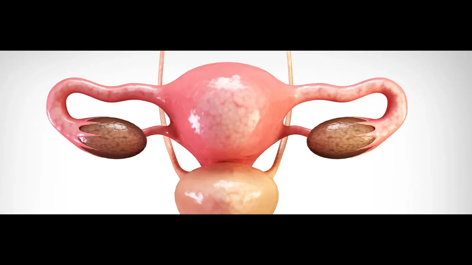
A uterine polyp—also called an endometrial polyp—is a localised overgrowth of endometrial glands and stroma forming a soft, vascular projection into the uterine cavity. It is not a fibroid.
Polyps can be single or multiple. Some attach by a slender stalk (pedunculated), while others have a broad base flush with the uterine wall (sessile). They range in size from a few millimetres to several centimetres across.
| Feature | Uterine (Endometrial) Polyp | Uterine Fibroid (Leiomyoma) |
|---|---|---|
| Origin | Endometrial mucosal lining | Muscular wall of the uterus (myometrium) |
| Texture | Soft, vascular, pliable tissue | Dense, firm fibrous-muscular tissue |
| Relation to Cavity | Grows directly inside the endometrial cavity | Only submucosal fibroids bulge into the cavity |
| Primary Treatment | Targeted hysteroscopic polypectomy | Hysteroscopy, laparoscopy, or observation |
This distinction matters in reproductive medicine. The endometrial cavity is where implantation takes place. A benign polyp may become a mechanical obstacle or alter the local environment if it occupies that space.
What causes endometrial polyps?
The precise reason why polyps develop is not always known. They are seen more often with increasing age and with some exposures, such as tamoxifen; they may also be found during infertility evaluation. These associations do not identify a single cause, and having a polyp is not a reflection of lifestyle or personal fault.
Recognised associations and clinical contexts include:
- Age: Polyps become more common with increasing age.
- Medication exposure: Tamoxifen is one recognised association.
- Infertility evaluation: Polyps may be found while investigating difficulty conceiving; this is a context in which they are detected, not necessarily a cause.
Many women diagnosed with polyps have no recognised association at all. They can also occur in young, healthy women trying to conceive, and their presence is not a reflection of lifestyle or personal fault.
Symptoms: subtle clues versus silent findings
Many uterine polyps are completely silent and produce no symptoms whatsoever. When symptoms do occur, bleeding changes are the most common sign.
Typical symptoms include:
- Intermenstrual bleeding: Light spotting or bleeding between expected menstrual cycles.
- Heavier menstrual bleeding (menorrhagia): Periods that last longer or feel noticeably heavier.
- Post-coital spotting: Light bleeding immediately following sexual intercourse.
- Postmenopausal bleeding: Any unexpected vaginal spotting after menopause, which always warrants prompt evaluation.
- Subfertility: Difficulty conceiving despite regular ovulation and clear fallopian tubes.
Pelvic pain is uncommon with polyps alone unless they are exceptionally large or prolapsing through the cervix. Because these bleeding symptoms overlap with other conditions—such as ovulatory disorders, adenomyosis, or fibroids—imaging and, when needed, visual assessment help establish the diagnosis.
Why polyps matter for fertility and implantation
Not every polyp prevents pregnancy, but an intracavitary polyp may interfere with embryo implantation.
A polyp can occupy space within the cavity, and biological effects on the local endometrium have also been proposed. These mechanisms help explain why a cavity-distorting lesion may matter, but they do not mean that every polyp prevents implantation.
The American Association of Gynecologic Laparoscopists (AAGL) lists infertility among the clinical settings in which endometrial polyps may be found, while abnormal bleeding remains the most common presentation.
The evidence is not uniform. The older retrospective study by Lass et al. found that small polyps did not lower pregnancy rates, although miscarriage was numerically higher. A later retrospective study by Nishioka et al. (2023) reported higher pregnancy and live-birth rates after hysteroscopic removal than after curettage. These findings can inform discussion of removing a cavity-distorting or symptomatic polyp in selected patients, but they do not prove that every small, asymptomatic polyp must be removed or that removal guarantees a live birth.
To learn more about preparing the lining for transfer, read our guide on endometrial thickness before embryo transfer.
How uterine polyps are diagnosed
Evaluation may involve one or more methods, depending on the symptoms and how clearly the uterine cavity needs to be seen:
- Transvaginal ultrasound (TVS): This is often the first test used. A polyp may appear as a focal hyperechoic thickening of the endometrial stripe.
- Saline Infusion Sonography (SIS): Sterile saline is gently infused into the uterine cavity during ultrasound. The fluid distends the walls, providing a crisp outline of the polyp’s size, base, and exact attachment site.
- Diagnostic Hysteroscopy: This examination provides a direct view of the uterine cavity. A slender camera (hysteroscope) is guided through the cervix into the cavity under fluid distension, allowing the physician to inspect the lining under direct magnification.
- Histopathological examination: Removed tissue is evaluated under a microscope to determine its nature and check for hyperplasia or atypical changes.
Diagnostic hysteroscopy provides a direct view of the cavity and can allow treatment during the same procedure when that is appropriate. Whether it is needed depends on the ultrasound findings, symptoms, and clinical plan. For more detail, see our guide on hysteroscopy in female infertility.
When to remove a polyp versus when to observe
Management is never one-size-fits-all. The decision between observation and removal depends on symptoms, size, location, age, menopausal status, and reproductive plans.
Watchful waiting (observation)
May be considered for:
- Small polyps (under 10 mm) discovered incidentally in premenopausal women without bleeding symptoms.
- Polyps situated away from the primary implantation zone when fertility treatment is not urgently planned.
- Small polyps that may regress spontaneously during subsequent menstrual cycles.
Hysteroscopic polypectomy (removal)
May be considered when:
- Abnormal uterine bleeding or intermenstrual spotting is present.
- Fertility treatment or IVF is planned and the polyp occupies or distorts the cavity.
- The patient is postmenopausal or the bleeding pattern requires tissue assessment.
- The lesion is large, multiple, persistent, or its nature is uncertain on imaging.
When removal is chosen, operative hysteroscopy uses direct visual guidance and usually avoids abdominal incisions. AAGL guidance does not favour blind removal when guided removal is available; the appropriate technique and anaesthetic plan depend on the patient and the procedure.
Dr. Aksoy’s Approach
The word “polyp” often causes more alarm than it deserves: most endometrial polyps are benign. For Dr. Senai Aksoy, the key question is whether the polyp occupies the part of the cavity where the embryo has to implant. A tiny polyp that causes no symptoms may simply be observed in some premenopausal patients. When a polyp causes abnormal bleeding, distorts the cavity or is found before an embryo transfer, targeted hysteroscopic removal may be discussed. Hysteroscopy lets the clinician see the cavity directly and, if the polyp is removed, send the tissue for examination.
Uterine polyps and IVF: timing the embryo transfer
In IVF and frozen embryo transfer (FET) cycles, assessing and, when appropriate, treating the uterine cavity is an important part of preparation.
When a polyp is diagnosed during ovarian stimulation or before transfer, one possible approach is to complete egg collection and fertilisation, freeze the resulting embryos, and perform hysteroscopic polypectomy in a later cycle. The choice depends on the polyp’s size and location, symptoms, treatment timing, and the overall clinical plan.
A 2026 retrospective cohort study found no difference in live-birth rates between fresh transfer and a freeze-all strategy followed by hysteroscopy. Because this was not a randomised trial, it supports individualised planning rather than a single rule for every patient.
There is no universally required waiting period after polypectomy. Timing is individualised according to healing, symptoms, the extent of the procedure, and the treatment plan.
Evaluating the cavity also involves assessing other factors that influence implantation, such as adenomyosis and IVF or co-existing fibroids and IVF.
Related reading
- Adenomyosis and IVF: Impact on Implantation
- Fibroids and IVF: When They Matter and When They Do Not
- Limits of Laparoscopic Myomectomy
- Hysteroscopy in Female Infertility: Clinical Evidence Guide
Frequently asked questions
Are uterine polyps cancerous?
Most endometrial polyps are benign. The risk of atypical or malignant change is higher with increasing age and postmenopausal bleeding, so tissue that is removed is usually sent for histopathological assessment.
Can a uterine polyp prevent natural pregnancy?
It may, particularly if it occupies the central or fundal cavity or obstructs a tubal opening. However, many women with small polyps conceive without difficulty, so the finding is interpreted in context.
Does every uterine polyp require surgical removal?
No. Small, asymptomatic polyps may regress spontaneously or simply be monitored in selected patients. Removal may be discussed when symptoms, abnormal bleeding, cavity distortion, or fertility plans make treatment relevant.
Is hysteroscopic polyp removal painful?
Hysteroscopic polypectomy may be performed with local anaesthesia, sedation, or another anaesthetic plan depending on the procedure and the patient. Pain and recovery vary; mild cramping and light spotting for a few days are common after the procedure.
Can uterine polyps grow back after removal?
Yes, recurrence is possible. Follow-up is guided by symptoms and the clinician’s assessment; an ultrasound may be appropriate if symptoms return.
What is the difference between a polyp and a fibroid?
A polyp arises from the endometrial mucosal lining and is soft and vascular. A fibroid (myoma) arises from the muscular uterine wall and is dense and firm. Both are usually benign, but they are evaluated and managed differently.
How soon after polyp removal can I attempt pregnancy or an embryo transfer?
There is no single interval that applies to everyone. The timing is individualised according to the extent of removal, healing, symptoms, and the planned treatment.
Sources
- AAGL Practice Report. Practice guidelines for the diagnosis and management of endometrial polyps. J Minim Invasive Gynecol. 2012;19(1):3-10.
- Lass A, Williams G, Abusheikha N, Brinsden P. The effect of endometrial polyps on outcomes of in vitro fertilization (IVF) cycles. J Assist Reprod Genet. 1999;16(8):410-415.
- Nishioka M, Maezawa T, Takeuchi H, et al. Pregnancy Rates after Hysteroscopic Endometrial Polypectomy versus Endometrial Curettage Polypectomy: A Retrospective Study. Medicina (Kaunas). 2023;59(10):1868.
- Bosteels J, van Wessel S, Weyers S, et al. Hysteroscopy for treating subfertility associated with intrauterine adhesions, endometrial polyps, submucous fibroids, or uterine septa. Cochrane Database Syst Rev. 2018;12(12):CD009461.
- Schutyser V, Agius MP, Drakopoulos P, et al. Managing suspected endometrial polyps during IVF/ICSI stimulation: retrospective cohort study of fresh versus freeze-all strategies. Reprod Biomed Online. 2026;53(1):105357.
Add as a Preferred Source on Google
You can add draksoyivf.com as one of your preferred health information sources on Google.

The content has been created by Dr. Senai Aksoy and medically approved.