
Uterine Fibroids and Fertility: Which Fibroids Matter Most
Key Takeaways
Not every fibroid affects fertility the same way; location is the decisive factor. Submucosal fibroids and cavity-distorting lesions are most likely to interfere with implantation and miscarriage risk, while subserosal fibroids usually do not. Whether to remove a fibroid depends on location, symptoms, age, ovarian reserve, and treatment timing.
Key evidence: ASRM guideline on myoma removal to improve fertility (2017) Systematic review: surgical treatment of fibroids for subfertility (Metwally et al., Cochrane 2012) Updated review: fibroids and infertility (Pritts et al., Fertil Steril 2009)
Uterine fibroids are extraordinarily common benign tumours arising from the smooth muscle wall of the uterus (myometrium). Up to 70% of women develop fibroids by age 50, and many discover them by coincidence during a routine pelvic ultrasound or when starting an initial fertility work-up.
When a scan reveals a fibroid, the first question almost every patient asks is: Does this need to come out before I try to conceive, or before we start IVF?
In most cases, the answer is no. Merely having a fibroid is rarely a reason for surgery. What matters fundamentally is where the fibroid sits relative to the uterine cavity, whether it distorts the endometrium where an embryo must implant, whether it causes symptoms, and how surgical recovery fits into your broader reproductive timeline.
What are uterine fibroids, and how are they classified?
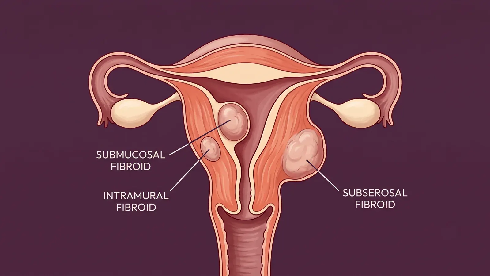
Fibroids (leiomyomas) vary widely in number, diameter, and position. For reproductive planning, gynecologists rely on the international FIGO classification system (types 0 to 8), which defines their anatomical relationship to the uterine lining:
- Submucosal (FIGO 0, 1, 2): These grow directly underneath or protrude into the endometrial cavity. Even a small 1 to 2 cm submucosal fibroid can mechanically obstruct embryo implantation or cause chronic local inflammation.
- Intramural (FIGO 3, 4, 5): These sit within the muscular wall. FIGO 3 touches the lining, FIGO 4 is entirely intramural, and FIGO 5 extends toward the outer surface. Large intramural fibroids can impinge upon or distort the cavity.
- Subserosal (FIGO 6, 7): These project outward from the external surface of the uterus. They leave the endometrial cavity undisturbed and rarely interfere with natural conception or embryo transfer.
Who is affected by this evaluation?
This assessment is especially important if you have been diagnosed with a fibroid and:
- You are preparing for an IVF or frozen embryo transfer (FET) cycle;
- You have experienced recurrent pregnancy loss or repeated implantation failures;
- You suffer from heavy, painful menstrual bleeding (menorrhagia) alongside fertility delays;
- Your routine scan noted an intramural fibroid approaching 4 cm or larger.
Accurate evaluation requires imaging that visualizes the uterine cavity with precision. A standard two-dimensional scan is often insufficient; three-dimensional transvaginal ultrasound, saline infusion sonohysterography (SIS), diagnostic hysteroscopy, or pelvic MRI are typically employed to establish whether the endometrial cavity is displaced.
How fibroids affect fertility and reproductive outcomes
Not all fibroids impact conception in the same way. The scientific literature demonstrates a clear biological hierarchy:
- Mechanical distortion of the cavity: Submucosal and large cavity-distorting intramural fibroids alter the architectural contour of the endometrium, disrupting the contact surface required for blastocyst apposition and invasion (Pritts et al., 2009).
- Altered endometrial receptivity: Fibroids adjacent to the endometrium trigger localized microvascular changes, persistent low-grade inflammation, and shifts in key implantation factors such as HOXA10 and leukemia inhibitory factor (Bulun, 2013).
- Disrupted uterine peristalsis: intramural lesions can perturb the coordinated muscular contractions that guide sperm migration and early blastocyst positioning.
- Tubal ostium occlusion: While less frequent, a large cornual fibroid can mechanically compress the interstitial portion of the fallopian tube.
In assisted reproduction, systematic reviews consistently demonstrate that removing a submucosal fibroid significantly restores clinical pregnancy and live birth rates. In contrast, removing an intramural fibroid that does not distort the cavity has not demonstrated a reliable, reproducible improvement in live birth outcomes (Metwally et al., Cochrane 2012 ; ASRM Practice Committee, 2017).
What the medical evidence proves — and what remains uncertain
High-quality evidence guides when surgery is beneficial and when it introduces unnecessary risks:
| Clinical Scenario | Strength of Evidence | Key Clinical Takeaway |
|---|---|---|
| Submucosal fibroids (FIGO 0, 1, 2) | Strong (Systematic reviews, ASRM guidelines) | Significantly lower pregnancy rates and double miscarriage risk. Hysteroscopic resection restores outcomes to near-normal. |
| Cavity-distorting intramural fibroids | Strong (Systematic reviews) | Impair implantation; surgical removal is generally recommended prior to embryo transfer. |
| Non-distorting intramural fibroids | Mixed / Uncertain (Conflicting cohort studies) | Modest reductions in IVF success reported primarily above ~4–5 cm. Routine surgical removal has not shown clear live-birth benefits in RCTs. |
| Subserosal fibroids (FIGO 6, 7) | Weak / No effect (Meta-analyses) | No measurable adverse effect on fertility or pregnancy rates. Surgery is reserved for severe pain or pressure symptoms. |
Because clinical trials vary in how strictly they define “cavity distortion,” reproductive specialists evaluate each patient individually rather than relying on an arbitrary size cutoff alone.
Evaluation and surgical management options
Treatment should always be tailored to the specific anatomical subtype, symptoms, and reproductive goals:
1. Hysteroscopic myomectomy
Hysteroscopy is the definitive minimally invasive treatment for intracavitary and submucosal fibroids (FIGO 0 and 1, and selected FIGO 2). Performed vaginally without abdominal incisions, it is an outpatient procedure associated with rapid recovery. Resection eliminates the intracavitary obstacle and directly restores the implantation bed (ASRM, 2017).
2. Laparoscopic myomectomy
For symptomatic subserosal fibroids or significant intramural lesions that distort the uterine contour, minimally invasive laparoscopic myomectomy offers precise enucleation and multilayered myometrial reconstruction. Surgical expertise is paramount: careful suturing is critical to prevent post-operative pelvic adhesions and ensure robust uterine wall integrity during a future pregnancy.
3. Open (laparotomic) myomectomy
When fibroids are exceptionally large (often exceeding 8 to 10 cm), highly numerous, or deeply embedded near major uterine blood vessels, laparotomy remains a safe, controlled approach. Although recovery takes longer (4 to 6 weeks), open access allows meticulous tactile palpation and multi-layer reconstruction.
4. Active observation (watchful waiting)
For asymptomatic intramural or subserosal fibroids that leave the cavity completely untouched, observation is frequently the wisest approach. In women over 35 or those with diminished ovarian reserve, avoiding a surgical delay of several months preserves invaluable reproductive time.
Comparing treatment paths at a glance
| Decision Factor | Hysteroscopic Myomectomy | Laparoscopic Myomectomy | Open Laparotomy | Active Observation |
|---|---|---|---|---|
| Submucosal fibroid (FIGO 0–1) | First-line gold standard | Rarely indicated | Not indicated | Only if patient declines surgery |
| Cavity-distorting intramural >4 cm | Possible if mostly cavitary | Common choice | Preferred if multiple or deep | Inadvisable before transfer |
| Large symptomatic subserosal | Not applicable | First choice | If laparoscopy unavailable | If symptoms are negligible |
| Patient age >38 or low AMH | Minimal delay (1–2 cycles) | Balance delay vs benefit | Avoid long delay | Strongest option if non-distorting |
| Timing to embryo transfer | ~4 to 6 weeks | ~3 to 6 months healing | ~3 to 6 months healing | Immediate cycle start |
Practical planning and the “freeze-all” strategy
For international patients travelling for IVF, reproductive timing must be planned strategically. If a cavity-distorting fibroid requires removal:
- Hysteroscopy: Typically requires a 2-to-3 day stay, followed by an ultrasound check 4 weeks later. Embryo transfer can often occur the following cycle.
- Laparoscopy or Laparotomy: Requires a 3-to-6 month uterine healing period before embryo transfer to ensure sound myometrial scar healing and minimize the risk of uterine rupture in pregnancy.
The “Freeze-All” Strategy for Women with Low Ovarian Reserve:
When a patient has diminished ovarian reserve or advanced maternal age, spending 6 months on surgery and recovery before egg retrieval can lead to a further drop in egg quality or quantity. In these cases, the optimal evidence-based strategy is to undergo ovarian stimulation and vitrify all blastocysts first (“freeze-all”). Once embryos are safely preserved in the laboratory, myomectomy can be performed without any pressure on the biological clock, followed by frozen embryo transfer after complete healing.
When to seek prompt medical advice
Most fibroids are slow-growing and benign. However, you should contact your physician promptly if you experience:
- Sudden, severe pelvic pain or acute localized tenderness (which may signal fibroid red degeneration or torsion of a pedunculated subserosal fibroid);
- Prolonged, heavy menstrual bleeding resulting in severe fatigue, dizziness, or anemia;
- Rapid uterine enlargement over a few months;
- Acute urinary retention or severe rectal pressure.
Elective fertility surgery should be deferred in the presence of untreated pelvic inflammatory disease, uncorrected coagulation disorders, or active pregnancy.
Dr. Aksoy’s Clinical Approach
The Perspective of Dr. Senai Aksoy
“In clinical consultations, the question I hear most often is not simply whether a fibroid is present, but where it sits. An embryo implants inside the endometrial cavity. Therefore, even a small 1.5 cm fibroid that breaches the cavity can impair pregnancy, whereas a 6 cm fibroid growing outward away from the lining may not affect embryo transfer at all.
The second major misconception is assuming that surgery always improves the odds. For an intramural fibroid that does not distort the cavity, myomectomy does not reliably increase live birth rates — and it can delay treatment by three to six months while introducing risks of surgical adhesions and uterine wall scarring. In a woman aged 38 or older, or someone with a low AMH, that delay often costs more in reproductive potential than the fibroid itself.
I frequently share this example: two women can both have a 4 cm fibroid on their ultrasound report. One has a submucosal component pushing the lining, which must be resected before transfer. The other has an outward subserosal growth that can be safely left alone. Same centimeter reading on paper — completely different reproductive implications.
What matters in clinical practice is looking beyond the ultrasound ruler. FIGO subclassification, distance to the endometrium, cavity distortion, symptoms, age, and ovarian reserve must all be weighed together. Our goal before IVF is never a theoretically pristine, fibroid-free uterus; our goal is a healthy, receptive cavity that allows your embryo to implant and grow.”
Key questions to discuss with your specialist
Before making a surgical decision, review these targeted questions with your reproductive endocrinologist:
- What is the exact FIGO classification of my fibroid, and is there any documented distortion of the endometrial lining on 3D ultrasound or hysteroscopy?
- Would you recommend removing this fibroid if I were not actively trying to conceive?
- How will the mandatory 3-to-6-month post-operative healing window affect my ovarian reserve and age-dependent fertility timeline?
- Would it be medically advantageous to perform egg retrieval and freeze embryos before any surgical intervention?
- What are the documented risks of intrauterine adhesions or myometrial scarring for my specific fibroid location?
Related reading
- Adenomyosis and IVF: When It Matters and How Treatment Is Tailored
- Limits of Laparoscopic Myomectomy: Safety and Fertility Considerations
- Uterine Polyps: Symptoms, Diagnosis, and When They Matter for Fertility
FAQ
Do all uterine fibroids reduce fertility?
No. Many women with uterine fibroids conceive naturally without difficulty. Impact on fertility is almost entirely governed by anatomical location. Subserosal fibroids rarely have any negative effect, whereas submucosal fibroids that distort the endometrial cavity reduce implantation rates and double miscarriage risk.
Should every fibroid be surgically removed before IVF?
No. Surgery is only warranted when a fibroid clearly distorts the endometrial cavity, causes severe symptoms like debilitating bleeding, or physically blocks access during egg retrieval. Routine removal of asymptomatic intramural fibroids that leave the cavity smooth does not improve live birth rates and introduces unnecessary surgical risks.
Can intramural fibroids matter even if they do not touch the cavity?
In selected cases, yes. Large intramural fibroids exceeding 4 to 5 cm may alter local blood flow, trigger low-grade inflammatory changes, or disturb uterine contractions even without clear cavity distortion. However, whether removing them improves IVF live birth rates remains debated in clinical trials, and the decision must balance surgical delay against maternal age and ovarian reserve.
Are subserosal fibroids generally harmless for pregnancy?
Yes. Because subserosal fibroids grow outward toward the abdominal cavity, they do not impinge upon the endometrium or impede embryo implantation. Surgical excision is reserved only for very large lesions that cause severe chronic pelvic pain, urinary frequency, or bowel compression.
Sources
- Practice Committee of the American Society for Reproductive Medicine. Removal of myomas in asymptomatic patients to improve fertility and/or reduce miscarriage rate: a guideline. Fertil Steril. 2017;108(3):416–425. doi:10.1016/j.fertnstert.2017.06.034.
- Metwally M, Cheong YC, Horne AW. Surgical treatment of fibroids for subfertility. Cochrane Database Syst Rev. 2012;(11):CD003857. doi:10.1002/14651858.CD003857.pub3.
- Pritts EA, Parker WH, Olive DL. Fibroids and infertility: an updated systematic review of the evidence. Fertil Steril. 2009;91(4):1215–1223. doi:10.1016/j.fertnstert.2008.01.051.
- Donnez J, Dolmans MM. Uterine fibroid management: from the present to the future. Hum Reprod Update. 2016;22(6):665–686. doi:10.1093/humupd/dmw023.
- Bulun SE. Uterine fibroids. N Engl J Med. 2013;369(14):1344–1355. doi:10.1056/NEJMra1209993.
Add as a Preferred Source on Google
You can add draksoyivf.com as one of your preferred health information sources on Google.

The content has been created by Dr. Senai Aksoy and medically approved.